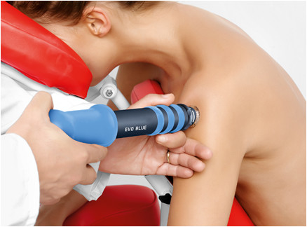
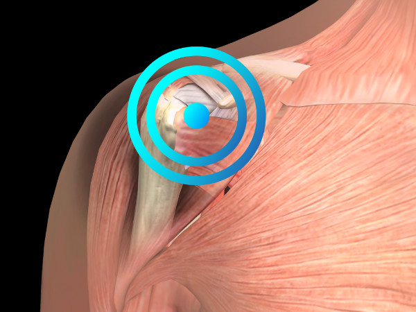
RADIAL PROTOCOL
Radial protocol values
Pressure: 2 bar
Impulses: 2,500
Frequency: 10–12
Applicator: 15 mm
Total energy flux density dose sum: 103 mJ/mm²
Number of sessions: 3 (1 per week)
Medical information
The term subacromial shoulder syndrome is commonly used synonymously with rotator cuff disease, rotator cuff tendinitis, and shoulder impingement syndrome.
As calcific tendinitis of the shoulder can also cause shoulder pain, subacromial syndrome may additionally include calcific shoulder tendinitis. In some cases, the term “rotator cuff tendinitis” is confused with shoulder bursitis; however, both terms refer to inflammation within a specific area of the shoulder joint (i.e., the subacromial space) that causes a common set of symptoms known as shoulder impingement syndrome (SIS). The term SIS is descriptive and refers to compression of the rotator cuff tendons and bursa between the bones (i.e., within the subacromial space). In most severe cases, SIS consists of a combination of inflammation of the rotator cuff tendons (tendinitis) and inflammation of the synovial bursa surrounding these tendons (bursitis). In many cases of SIS, the subacromial space becomes reduced due to anatomical changes in the bone structure compared to that of a healthy individual.
The condition is usually caused by an initial injury that triggers the inflammatory process. This may lead to thickening of the tendons or the bursa, causing these structures to occupy more space and become further compressed, which in turn increases inflammation. As a result, the condition may become self-perpetuating, leading to a vicious cycle of inflammation, tendon and bursa thickening, impingement of these structures, further inflammation, and so on.
This condition is diagnosed based on its clinical symptoms. Imaging studies should be used to rule out other causes of shoulder pain or to confirm the diagnosis of SIS when there is diagnostic uncertainty.
SIS is the most common cause of shoulder pain, and repetitive activities or movements involving lifting the arms above shoulder level at work or during sports practice — including swimming, throwing sports, tennis, weightlifting, golf, volleyball, and gymnastics — represent the main risk factors for developing SIS. The likelihood of developing the condition also increases with age.
Regarding treatment, three distinct stages of SIS are generally recognised:
- Phase 1 (acute inflammation, oedema, and haemorrhage within the rotator cuff): should be managed with conservative measures such as rest, ice application, physiotherapy, and non-steroidal anti-inflammatory drugs (NSAIDs);
- Phase 2 (prolongation of Phase 1, with progression to fibrosis and rotator cuff tendinitis): may be managed with conservative treatment, radial shock wave therapy (rESWT®), or surgery if conservative treatment and rESWT® fail;
- Phase 3 (mechanical rupture of the rotator cuff tendon and/or structural changes in the coracoacromial arch with osteophytes along the anterior acromion): surgery is considered the only effective treatment option.
STUDIES
Engebretsen K, Grotle M, Bautz-Holter E, et al. Radial extracorporeal shock wave treatment compared with supervised exercises in patients with subacromial pain syndrome: a single blind randomised study. Brit Med J 2009; 339:b3360. Refer to the study.
FOCAL PROTOCOL
Focal protocol values
Penetration depth: 20 mm
Total energy flux density per shock wave emission: 0.138–0.22 mJ/mm²
Number of sessions: 3 sessions (1 per week)
Frequency (Hz): 8 Hz
Impulses: 2,000–2,500