Primary and secondary lymphedema
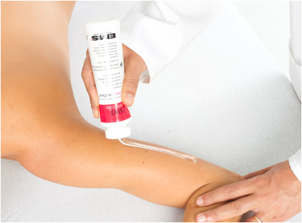
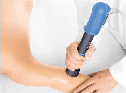
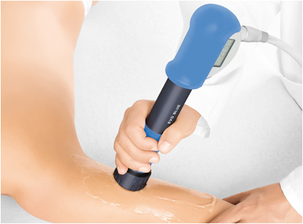
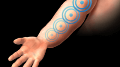
RADIAL PROTOCOL
Radial protocol values
Pressure: 3 bar
Impulses: 1,000
Frequency: 5–10 Hz
Applicator: 36 mm
Total energy flux density dose: 23 mJ/mm²
Number of sessions: 10 (2 per week)
Medical Information
Lymphedema may be primary or secondary. Primary lymphedema is a lymphatic malformation that develops during the final stage of lymphangiogenesis. In contrast, secondary lymphedema results from disruption or obstruction of the lymphatic system.
Secondary lymphedema may occur as a consequence of tumors, surgical procedures, infections, inflammation, radiotherapy treatments, and trauma. It is one of the most significant complications following surgical treatment for breast cancer and has a considerable impact on quality of life. A substantial number of women develop secondary lymphedema after breast cancer removal surgery, with an incidence ranging from 6% to 63% depending on the population studied, the measurement parameters used, and the duration of follow-up.
Lymphedema is divided into three stages:
- Stage IA (latent lymphedema) presents without clinical evidence of edema, but with impaired lymphatic transport capacity.
- Stage IB (early lymphedema) is characterized by edema that is totally or partially reduced with rest and drainage.
- In stage IIA (progressive lymphedema), the lymphatic transport capacity disappears and fibro-indurative skin changes begin to develop.
- Stage IIB (fibrotic lymphedema with column-shaped limbs) presents with lymphostatic skin changes and worsening disability.
- Stage IIIA (elephantiasis) is characterized by scleroindurative pachydermatitis and papillomatous lymphostatic verrucosis, together with life-threatening disability.
- Stage IIIB consists of extreme elephantiasis with complete disability.
The diagnosis of lymphedema is based on the clinical characteristics of the disease (measurement of limb circumference before and after surgery, where a difference greater than 2 cm indicates the development of lymphedema). Imaging studies (plain radiographs, duplex ultrasound, isotopic lymphoscintigraphy, and other imaging modalities) should be considered to rule out other causes of increased limb circumference or to confirm the diagnosis of lymphedema when uncertainty exists.
Treatment should begin with manual lymphatic drainage and complete decongestive therapy focused on compression bandaging. An alternative approach is sequential intermittent pneumatic compression using pump devices. Radial shock wave therapy (rESWT®) has demonstrated effectiveness in the treatment of stage IIA and IIB lymphedema. Surgery should be considered in recalcitrant cases of lymphedema that do not respond or respond poorly to the aforementioned treatment options. Surgical options include lymphovenous or lymphatico-venous-lymphatic bypass anastomosis, segmental lymphatic interposition, free lymph node transfer, and ablative surgery in cases of massive limb changes or fibrotic induration.
STUDIES
Michelini S, Failla A, Moneta G, et al. Treatment of primary and secondary lymphedema with shockwave therapy. Eur J lymphol 2008; 19:10.