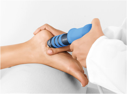
RADIAL PROTOCOL
Radial protocol values
Pressure: 2.5 bar
Pulses: 2,500
Frequency: 10–12 Hz
Applicator: 15 mm
Total energy flux density dose sum: 166 mJ/mm²
Number of sessions: 3 (1 per week)
Medical information
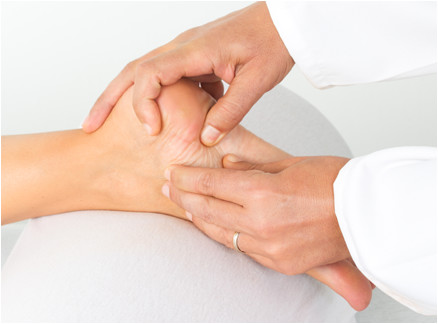
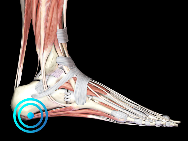
Plantar fasciopathy (PF) is an acute or chronic painful disorder of the plantar fascia, which extends between the medial tuberosity of the calcaneus and the proximal phalanges of the toes.
It is the most common cause of plantar heel pain, accounting for approximately 11–15% of medical consultations related to painful foot symptoms. Its main clinical symptom is heel pain, especially in the morning or after a period of rest. Patients often report pain relief after walking. The origin of the pain is generally located in the plantar fascia, specifically at the medial calcaneal tubercle.
Passive dorsiflexion of the toes may aggravate pain in some patients, particularly in those with chronic PF. Patients suffering from chronic PF may also present inflammation of the heel pad. This condition is diagnosed based on its clinical symptoms.
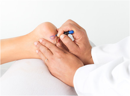
Diagnostic imaging should be used to rule out other causes of plantar heel pain or to confirm the diagnosis of PF when there is uncertainty. However, histological examination of tissue samples (biopsies) obtained from patients during surgery for chronic symptoms has shown that chronic PF is associated with degenerative changes in the fascia. Consequently, the condition is more accurately described as “fasciopathy” rather than “fasciitis”, and it resembles the pathological process observed in other overuse tendon disorders.
In the United States, more than two million people are treated for PF each year. Throughout their lifetime, up to 10% of the population will experience plantar heel pain. Both athletes and elderly individuals commonly seek medical attention for PF.
Treatment of the condition should begin with conservative management approaches such as rest, physiotherapy, stretching, exercise programs, shoe insoles, orthotics, night splints, non-steroidal anti-inflammatory drugs, and local corticosteroid injections. Patients who do not respond to conservative treatment within six months (approximately 10–20% of patients) may be considered for radial shock wave therapy (RSWT®). In the most persistent cases of PF, surgery should also be considered.
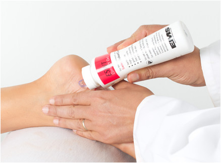
During shock wave therapy treatment (focused or radial), the current consensus recommends avoiding any anti-inflammatory medication or methods that may reduce vascularization in the treated area, such as the application of ice. In the case of infiltrations/injections, it is recommended to postpone shock wave therapy treatment for at least four weeks after the last injection.
STUDIES
Gerdesmeyer L, Frey C, Vester J, et al. Radial extracorporeal shock wave therapy is safe and effective in the treatment of chronic recalcitrant plantar fasciitis: results of a confirmatory randomized placebo-controlled multicenter study. Am J Sports Med 2008;36:2100-2109. Refer to the study.
Ibrahim M, Donatelli R, Schmitz C, et al. Successful treatment of chronic plantar fasciitis with two sessions of radial extracorporeal shock wave therapy. Foot & Ankle Int. 2010 May; 31 (5):391-97 20460065. Refer to the study.
FOCAL PROTOCOL
Focal protocol values
Penetration depth: 10–20 mm
Total energy flux density per shock wave emission: 0.077–0.355 mJ/mm²
Number of sessions: 3–5 (1–2 per week)
Frequency: 8 Hz
Pulses: 2,500–3,000
Medical information
Plantar fasciitis or heel spur is a common condition. Plantar heel pain occurs when excessive tension is applied to the insertion of the plantar fascia and certain foot muscles at the distal calcaneus. The precise etiology of plantar fasciitis has not yet been fully elucidated. It has been suggested that the condition may have a multifactorial origin, resulting from repetitive mechanical overload at the origin of the plantar aponeurosis. This leads to repeated microtrauma of the plantar fascia followed by tissue inflammation. Imaging studies often reveal a bony spur (heel spur). Patients initially complain of intermittent pain that gradually increases over time and significantly reduces the load-bearing capacity of the heel and sole of the foot.
Characteristically, the pain is most intense immediately upon getting out of bed in the morning, during weight-bearing activities, or after a prolonged period without pressure being applied to the heel; this pain generally decreases or disappears after taking a few steps.
The pain has been described as severe, sharp, and stabbing. It occurs during physical activity and often decreases when the foot is at rest. Treatment of plantar fasciitis with focused piezoelectric shock waves has become an established ESWT therapy.
STUDIES
Aqil A, Siddiqui MR, Solan M, et al. Extracorporeal shock wave therapy is effective in treating chronic plantar fasciitis: a meta-analysis of RCTs. Clin Orthop Relat Res 2013; 471:3645-52.
Chang KV et al. Comparative effectiveness of focused shock wave therapy of different intensity levels and radial shock wave therapy for treating plantar fasciitis: a systematic review and network meta-analysis. Arch Phys Med Rehabil. 2012 Jul;93(7):1259-68.
Gerdesmeyer L, Frey C, Vester J, et al. Radial extracorporeal shock wave therapy is safe and effective in the treatment of chronic recalcitrant plantar fasciitis: results of a confirmatory randomized placebo-controlled multicenter study. Am J Sports Med 2008; 36:2100-9.
Gollwitzer H et al. Clinically relevant effectiveness of focused extracorporeal shock wave therapy in the treatment of chronic plantar fasciitis: a randomized, controlled multicenter study.J Bone Joint Surg Am. 2015 May 6;97(9):701-8.
Haake M, Buch M, Schoellner C, et al. Extracorporeal shock wave therapy for plantar fasciitis: randomized controlled multicenter trial. BMJ 2003; 327:75.
Ibrahim MI et al. Long-term results of radial extracorporeal shock wave treatment for chronic plantar fasciopathy: A prospective, randomized, placebo-controlled trial with two years follow-up. J Orthop Res. 2017 Jul;35(7):1532-1538.
Njawaya MM et al. Ultrasound Guidance Does Not Improve the Results of Shock Wave for Plantar Fasciitis or Calcific Achilles Tendinopathy: A Randomized Control Trial. Clin J Sport Med. 2018 Jan;28(1):21-27.
Roerdink RL et al. Complications of extracorporeal shockwave therapy in plantar fasciitis: systematic review. Int J Surg. 2017 Oct; 46:133-145.
Rompe JD et al. Radial shock wave treatment alone is less efficient than radial shock wave treatment combined with tissue-specific plantar fascia-stretching in patients with chronic plantar heel pain. Int J Surg. 2015 Dec;24(Pt B):135-42.
Rompe JD, Meurer A, Nafe B, Hofmann A, Gerdesmeyer L. Repetitive low-energy shock wave application without local anesthesia is more efficient than repetitive low-energy shock wave application with local anesthesia in the treatment of chronic plantar fasciitis. J Orthop Res 2005; 23(4): 931-941
Saxena A et al. Treatment of Plantar Fasciitis with Radial Soundwave "Early" Is Better Than After 6 Months: A Pilot Study. J Foot Ankle Surg. 2017 Sep - Oct;56(5):950-953.
Speed CA, Nichols D, Wies J, et al. Extracorporeal shock wave therapy for plantar fasciitis. A double blind randomized controlled trial. J Orthop Res 2003; 21:937-40.
Sun J et al. Extracorporeal shock wave therapy is effective in treating chronic plantar fasciitis: A meta-analysis of RCTs. Medicine (Baltimore). 2017 Apr;96(15).
Johnson LG et al. Efficacy of Platelet-Rich Plasma in Soft Tissue Foot and Ankle Pathology. JBJS Rev 2022 Oct 3;10(10).
Naterstad IF et al. Efficacy of low-level laser therapy in patients with lower extremity tendinopathy or plantar fasciitis: systematic review and meta-analysis of randomized controlled trials. BMJ Open. 2022 Sep 28;12(9):e059479.
Lai Wei-Fu et al. The effectiveness of dextrose prolotherapy in plantar fasciitis: A systemic review and meta-analysis. Medicine (Baltimore) . 2021 Dec 23;100(51):e28216.