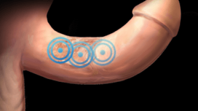
RADIAL PROTOCOL
Radial protocol values
Pressure: 2 bar
Impulses: 2000
Frequency (Hz): 10–12 Hz
Applicator (mm): 15 mm
Total energy flux density dose sum: 103 mJ/mm²
Number of sessions: 3 (1 per week)
Medical information
Peyronie’s disease, also known as induratio penis plastica, is an acquired penile curvature that may cause erectile dysfunction. When present, pain typically occurs during the early stage of disease progression and tends to decrease over time.
This condition is diagnosed based on its clinical symptoms. At the microscopic level, the disease is characterized by the formation of collagen and fibrin deposits, along with a reduction in elastic fibers, resulting in plaque formation within the tunica albuginea. This penile scarring leads to curvature during erection.
Peyronie’s disease has been proposed to result from abnormal wound healing, with the formation of collagen and fibrin deposits potentially caused by microtrauma. This hypothesis is supported by the finding that soft tissue injury may increase levels of transforming growth factor β1 (TGF-β1), which could potentially trigger a pro-inflammatory and pro-fibrotic cascade, followed by collagen deposition. However, no unified criteria currently exist regarding the pathophysiology of the disease process.
The incidence within the male population is approximately 1–3%, with peak incidence occurring between 40 and 70 years of age. Between 20% and 40% of men affected by Peyronie’s disease experience erectile dysfunction, and the condition is estimated to have significant psychological effects in up to 77% of affected individuals. Notably, the number of patients diagnosed with Peyronie’s disease appears to have increased since the introduction of oral sildenafil (Viagra).
Due to the lack of unified criteria regarding the pathophysiology of the disease, therapeutic approaches have not consistently provided reliable outcomes or complete symptom resolution. Current treatment strategies are aimed at modulating certain components of the wound-healing process. These include oral supplementation with vitamin E, omega-3 fatty acids, and the antioxidant coenzyme Q10, as well as pentoxifylline (a non-specific phosphodiesterase inhibitor with antifibrotic and anti-inflammatory properties). Other therapeutic strategies include topical application of verapamil, trifluoperazine, or magnesium sulfate, iontophoresis, intralesional injection of corticosteroids, collagenase, verapamil, or interferon alpha, radiotherapy, and penile traction therapy.
More recently, radial shock wave therapy (RSWT) has demonstrated efficacy in the treatment of Peyronie’s disease. The specific molecular and cellular mechanisms underlying the effects of RSWT in Peyronie’s disease are not yet fully understood, although they are reasonably believed to be related to the well-documented beneficial effects of RSWT on wound healing.
STUDIES
Palmieri A, Imbimbo C, Longo n, et al. A first prospective, randomized, double-blind, placebo-controlled clinical trial evaluating extracorporeal shock wave therapy for the treatment of Peyronie’s disease. Eur urol 2009; 56: 363-369. Refer to the study.
FOCAL PROTOCOL
Focal protocol values
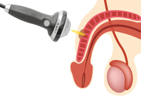
Treatment with the PiezoWave 2 device:
A total of 5000 pulses delivered at an energy flux density ranging from 0.16 mJ/mm² to 0.702 mJ/mm², depending on patient tolerance, and at a penetration depth of 0 mm.
The total 5000 pulses are distributed as follows:
- 2500 impulses using the G2 linear applicator along the corpora cavernosa at 0.16 mJ/mm².
- If a G4 or G6 applicator is available, 2500 pulses are delivered at 0.7 mJ/mm², depending on patient tolerance, and distributed across the plaque. If a focal applicator is not available, the full 5000 pulses are delivered using the linear applicator.
Adjunctive treatment: Tadalafil 5 mg, once daily dosage.
Medical information
To date, the available therapeutic options have not been particularly convincing. When treatment attempts with medication, including injections, were unsuccessful, the only remaining option was costly and sometimes complex surgical intervention.
With the introduction of shock wave therapy, a treatment modality is now available that may be used as an alternative to previous therapeutic approaches.
Initial studies have shown that ESWT can achieve a significant reduction in pain after only a few sessions and may help prevent disease progression. In addition, anecdotal user experiences have suggested that a degree of curvature correction may also be achieved.
Calcifications are frequently found within the so-called plaques, which are treated directly with ESWT. This approach may help reduce painful inflammatory processes, slow disease progression, and promote regeneration of erectile tissue.
STUDIES
C. Ratz, C.H.R. Ratz, S. Thieme-Martens, 654 Long-term Results of Patient Satisfaction After Low-intensity Shockwave Treatment (Li ESWT) of Erectile Dysfunction and Peyronie's Disease in an Urological Private Practice, The Journal of Sexual Medicine, Volume 15, Issue Supplement_3, July 2018, Page S378, https://doi.org/10.1016/j.jsxm.2018.04.561
Kuehhas FE, Weibl P, Georgi T, Djakovic N, Herwig R. Peyronie's Disease: Nonsurgical Therapy Options. Rev Urol. 2011;13(3):139-46. PMID: 22110397; PMCID: PMC3221554.
Briganti, A., Salonia, A., Zanni, G. et al. A Critical assessment of extracorporeal shock-wave therapy for Peyronie’s disease. Curr sex health rep 1, 19–23 (2004). https://doi.org/10.1007/s11930-004-0014-3
Abdessater, Maher; Akakpo, William; Kanbar, Anthony; Parra, Jérome; Seisen, Thomas; Chartier-Kastler, Emmanuel; Drouin, Sarah J; Roupret, Morgan. Low-intensity extracorporeal shock wave therapy for Peyronie's disease: a single-center experience. Asian Journal of Andrology 24(1):p 45-49, Jan–Feb 2022. | DOI: 10.4103/aja.aja_40_21